#10 Pneumothorax & Pleural Disease

Whether you work in the ICU, on the medicine wards or in the prehospital setting, you've got to be familiar with the unsung hero of the thorax - the pleura! On this episode of Critical Care Time, Cyrus & Nick extol the virtues of this humble structure, cover a little comparative physiology and spend the latter half of their chat discussing all things pneumothorax. Sit back, relax and enjoy this episode packed with pulmonary and critical care pearls that will take your breath away!
Quick Take Home Points:
The pleura is made up of two layers - the viscera and parietal - that play different roles within the thorax
Pleural fluid serves two major roles: it allows the two layers of the pleura to slide easily during respiration, and increases surface tension between visceral and parietal pleura - keeping the layers adhered to one another
Pneumothorax can occur spontaneously for no obvious reason (primary) or in the setting of known lung disease (secondary) or they may be traumatic/iatrogenic (non-spontaneous)
A pneumothorax can be acutely life threatening or completely asymptomatic and, of course, management depends on the severity
A tension pneumothorax - a medical emergency - results when air accumulates in the pleural space to the point where the pressure in that hemithorax is such that it results in obstructive shock and ultimately death if air is not rapidly removed
Conservative management strategies for asymptomatic or very mildly symptomatic pneumothoracies include observation or observation with nitrogen washout
In more symptomatic cases, drainage via needle, needle/catheter or thoracostomy tube with an ambulatory valve can be considered
In moderately symptomatic cases, we recommend small bore thoracostomy tube placement with inpatient observation to ensure resolution, followed by appropriate follow up based upon the history
Pleurodesis (medical or surgical) can be considered in certain cases - specifically a a second, primary spontaneous pneumothorax or a first secondary spontaneous pneumothorax
Infographic:
Animated Infographic depicting the POCUS findings in pneumothorax.
Show Notes:
Pleural anatomy
Two layers: visceral pleural & parietal pleura
Visceral pleura: adhered to the viscera - in this case the lungs (and bronchi, nerves, blood vessels etc.)
Parietal pleura: adhered to the chest wall
Pleural invaginations form the fissures between the lobes of the lungs
Pleural fluid is produced by the pleura and exists within the pleural space - we don’t know exactly how this works - but theories suggest pleural fluid is derived from pleural microvessels with the parietal pleura *probably* playing the primary role
The parietal pleura is principally responsible for pleural fluid drainage owing to the lymphatic stomata seen on the parietal pleura
Pleural fluid resorption can increase 20-25x relative to steady state, in the event of pathologically increased levels of pleural fluid
The transpulmonary pressure = alveolar pressure - intrapleural pressure, should be positive because plural pressure should be negative, creating a “vacuum” effect that keeps the pleura adhered
Analogy: Think about a microscope slide. The liquid allows the cover slip to slide back and forth. This is exactly how the parietal and visceral pleura remain adherent and allow movement.
Perturbations can alter respiratory mechanics
Pleural manometry - either via a esophageal balloon catheter or via direct manometry during thoracentesis - can be used to assess the intrapleural pressure
Under normal circumstances, we have two distinct pleural cavities - unlike buffalos that share one pleural space - this means that violation of one space does not necessarily doom us to sudden onset, catastrophic, obstructive shock

The sliding of the two layers of the pleura is like a microscope coverslip on a glass slide.
What is a pneumothorax?
Pneumothorax = Air in the pleural space
Air can enter from “outside-in” (e.g. a sucking chest wound) or “inside-out” (e.g. a misplaced subclavian central line that violates the lung/small bronchi)
Spontaneous pneumothorax: no clear, inciting event - ruptured bleb
Non-spontaneous pneumothorax: clear, inciting event - trauma, iatrogenesis
Note: Pneumothoraces in patients on positive pressure is a big deal because these patients are receiving breaths under pressure that will naturally cause air to follow the path of least resistance - through a defect and into the pleural space; this can rapidly lead to tension pneumothorax.
Primary Spontaneous Pneumothorax: No known or apparent pre-existing lung disease
Numerous theories exist as to why this happens but generally speaking, subpleural blebs - not previously seen/diagnosed - rupture under certain conditions leading to the pneumothorax
Secondary Spontaneous Pneumothorax: Patient has pre-existing lung disease, for example:
COPD/A1AT Deficiency
Cystic Fibrosis
Malignancies - either primary pulmonary or metastatic disease
Infections - for example, PCP pneumonia
Cystic lung diseases such as DIP, LIP, LAM or Bert-Hogg-Dube
Underlying architectural disease usually due to connective tissue disease
Catamenial pneumothorax
Pneumothorax can cause a spectrum of disease from totally asymptomatic to obstructive shock & cardiac arrest
The latter is a function of tension physiology → tension pneumothorax
Pleural air accumulates to the point where the affected hemithorax - under pressure - exerts force on the great vessels & heart, impairing venous return and cardiac output, leading to profound shock and death if left untreated
High risk characteristics of a pneumothorax
Hemodynamic compromise
Significant hypoxia
Bilateral pneumothorax
Underlying lung disease
Age >50 with a significant smoking history
Hemopneumothorax
Size Criteria for a Pneumothorax
BTS: large = the presence of a visible rim of >2 cm between the lung margin and the chest wall (at the level of the hilum)
Not stated explicitly in 2023 guidelines, suggested but mentioned explicitly in prior guideline (2010)
2023 guideline focuses more on symptoms/presentation rather than size as a criteria that dedicates management
CHEST: large = >/= 3 cm apex-to-cupola distance
Diagnosis of Pneumothorax
Precipitous tension pneumothorax is a clinical diagnosis
Chest X-Ray (CXR) is commonly done and allows for size estimation
CT is not recommended as the first initial test as it is more time consuming that other options and a patient may decompensate in the scanner if they are tenuous
The better use of CT is to assess a patient who has a history of pneumothorax that has been or is being managed actively, for whom a reason for the pneumothorax is still being sought after
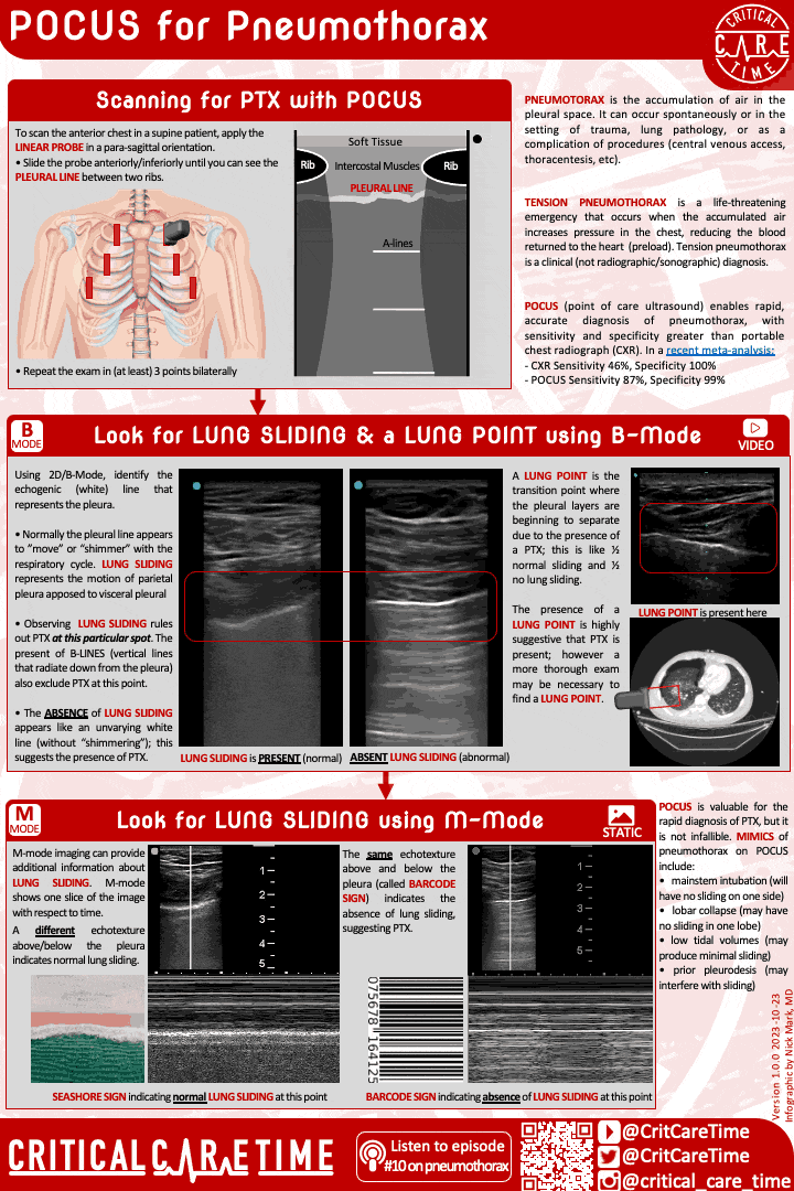
Point of Care Ultrasound (POCUS) for Pnuemothora: very effective and easy/quick!
2D Mode (aka B-mode): normal is the “ants marching” appearance where near the probe you should see intermittent, small amplitude movement “back and forth” indicative of the pleura sliding
A lung point is definition as the aforementioned that comes to an abrupt stop, highly suggestive of a pneumothorax at that particular location
The presence of B-Lines - the shimmering comet tails that emanate from the pleura, typically indicative of some degree of fluid in the alveoli - is highly suggestive of no pneumothorax at that particular location because pleural sliding is necessary for the formation of B-Lines on ultrasound
M-Mode (aka motion mode): allows for the assessment of pleural motion along a single line
Seashore sign / sandy beach: in M-Mode, there is a change in echotexture above/below the pleura. This looks like a seashore, where the waves and sand have very different textures. Seen under normal conditions.
Barcode sign or stratosphere sign: Pleural space looks the same above and below the pleura - just a bunch of horizontal lines throughout the ultrasonographic window - due to the entrainment of air in the pleural space and consequent artifact. Not normal, suggestive of pneumothorax.
Ultrasound is great but not a be-all-and-end-all
Your ultrasound could mislead you into thinking a patient has an acute pneumothorax in certain settings
Prior pleurodesis
Mainstem intubation if ultrasounding the contralateral hemithorax
Dense ARDS with significant fibrosis + low tidal-volume ventilation
Management of Pneumothorax
If shock is present → pleural evacuation, conservative/expectant management has no place in these patients. Several options exist:
Needle decompression: can be done in a pinch, limits options for further management
Needle with catheter: slightly better as catheter can allow for continued efflux of air, attachment to a 3-way stopcock for manual aspiration
Small bore, easily “gummed” up, easily kinked, may not allow for wire passage
Needle → wire → thoracostomy tube (12-14 Fr “Pigtail”): More definitive and secure management, allows for continuous suction of needed, can be hooked up to a pleural evacuation chamber
Our preferred method
Large-bore / surgical chest tube: Will get the job done, but at a cost to the patient with a higher risk of complications (laceration of intercostal vessels) without a benefit in most cases
Good option in a pinch, but no no faster compared to an operator well-trained in the use of a small bore thoracostomy tube placed via Seldinger technique
Asymptomatic patient
Asymptomatic patients with incidentally found primary spontaneous pneumothoraces can be managed conservatively
We recommend - in keeping with most expert opinion - obtaining an initial CXR followed by a repeat in 4-6 hours and another one in 24-48 hours, with appropriate outpatient follow up
This assumed good access to care and reliability
If secondary spontaneous pneumothorax - patient should be assessed for inpatient care and likely admitted for observation, given the presence of underlying lung disease which confers a higher risk for complication(s) / worsening
Minimally symptomatic patient
Nitrogen washout
Not recommended by guidelines at this time, but works theoretically & seen retrospectively by increasing the nitrogen gradient between air in the pleural space and arterial blood, thereby hastening reabsorption of pleural air
Our approach: If you are observation a patient anyway, it is a reasonable, very low risk “intervention” that can be trialed to hasten recovery
15 LPM NRB x4-6 hours followed by repeat CXR
Needle aspiration
Can insert a needle, aspirate air, and remove the needle, then image immediately after and again in 4-6 hours
Does not allow for continued management if pneumothorax reaccumulates
Needle aspiration with catheter
Allows for continued efflux of air, in theory
Could allow for entrainment of air from the environment (in theory)
Can kink or get “gummed” up
Our bottom line: For most patients in this category, observation with nitrogen washout x4-6 hours, followed by thoracostomy tube if necessary is the most pragmatic approach
One option from here in a patient with a thoracostomy tube can be the use of a Heimlich Valve - a one way valve that allows for continued efflux of air in the ambulatory setting
In theory this allows for safe discharge of the patient, assuming good/close follow up
In practice, pleural fluid can get trapped in the valve causing the valve to fail, creating a closed symptom and defeating the purpose of the valve in the first place
Moderately symptomatic patient
Small bore thoracostomy tube with pleural evacuation chamber
Minimizes pain, technically a straight forward procedure
Our practice:
Insert the thoracostomy tube based on the anatomy of the pneumothorax and patient factors, can consider lateral or anterolateral - or event anterior - placement to maximize comfort after scouting with ultrasound
Suction: We recommend brief/temporary suction to evacuate air that does not immediately efflux with needle placement. We usually would not recommend long term, continuous suction as this is not necessary for a simple pneumothorax and runs the risk of “formalizing” a tract and leading to a pesky bronchopleural fistula that needs more intensive management
Once stabilized, place the patient on water seal and get a CXR
Water seal means that air can escape the patient through the tube and through the water seal, but the seal will not allow air to entrain
We will generally have a patient on water seal for 12-24 hours with serial assessments for the presence of air leak(s) or other complicating factors
After the water seal has been in place and no further air bubbles are seen, another CXR is reasonable to confirm resolution of the pneumothorax
Practices differ at this point: It may be reasonable to remove the thoracostomy tube and have the patient follow up with their primary care provider
When possible, we like the added comfort/security of a “clamping trial” whereby the thoracostomy tube is physically clamped so that no air can escape
This is colloquially referred to as a “trial of life”: If the patient still had a subtle air leak, that may result in accumulation of air and symptoms which would change management versus true resolution of the pneumothorax
After a successful clamping trial, the tube can be removed and an occlusive dressing (with petroleum jelly / vaseline / xeroform or similar) can be placed +/- a stitch if necessary (usually a stitch is not required)
A word on pleurodesis
A patient with a first spontaneous pneumothorax deserves further work up, but management beyond resolving the pneumothorax is not necessary
In patients with a secondary pneumothorax or those with a second spontaneous, pleurodesis should be considered - elimination of the pleural space via surgical or medical means
This is usually a multi-disciplinary discussion between patient, pulmonary medicine/interventional pulmonary and possibly cardiothoracic surgery
Audio
Video
-
Lai-Fook SJ. Pleural mechanics and fluid exchange. Physiol Rev. 2004;84(2):385-410.
Charalampidis C, Youroukou A, Lazaridis G, et al. Physiology of the pleural space. J Thorac Dis. 2015;7(Suppl 1):S33-S37.
Grabczak EM, Krenke R, Zielinska-Krawczyk M, Light RW. Pleural manometry in patients with pleural diseases - the usefulness in clinical practice. Respir Med. 2018;145:230-236.
Baumann MH, Strange C, Heffner JE, et al. Management of spontaneous pneumothorax: an american college of chest physicians delphi consensus statement. Chest. 2001;119(2):590-602
Roberts ME, Rahman NM, Maskell NA, et al. British thoracic society guideline for pleural disease. Thorax. 2023;78(Suppl 3):s1-s42.
Grasmuk-Siegl E, Valipour A. “Nitrogen wash-out” in non-hypoxaemic patients with spontaneous pneumothorax: a narrative review. J Clin Med. 2023;12(13):4300.
-
-